

Specimen No. 3267 (Femur)
Artist
William Abraham Bell
(British, 1842 - 1920)
CultureAmerican
Dateca. 1865
MediumAlbumen print
DimensionsOverall, Image: 8 1/2 × 6 9/16 in. (21.6 × 16.7 cm)
Overall, Support: 13 5/8 × 10 3/4 in. (34.6 × 27.3 cm)
Overall, Mat: 16 1/16 × 20 in. (40.8 × 50.8 cm)
Overall, Support: 13 5/8 × 10 3/4 in. (34.6 × 27.3 cm)
Overall, Mat: 16 1/16 × 20 in. (40.8 × 50.8 cm)
InscribedThe number "3267" appears in white on the image on the front of the wooden pedestal on which the bone rests. The following is printed on the reverse of the print: "Surgeon General's Office. / ARMY MEDICAL MUSEUM. / SPECIMEN No. 3267. / Lower Half of Left Femur, exhibiting a / partially Consolidated Gunshot Fracture of the Shaft. The / deformed Callus encloses several Necrosed Splinters and a / battered Musket Ball. / Sergeant Sewell T. Douglas, Co. G, 1st Regiment Maine Heavy / Artillery, aged 28 years, was wounded at the battle of Spottsylvania, and / was admitted to Emory Hospital at Washington, May 22d. 1864. A / musket ball entering posteriorly, had fractured the lower third of the / left femur and lodged in the medullary cavity. / The injured limb was placed on a double inclined plane, and moderate extension was used. Internally, stimulants and tonics were employed. / In August, 1864, the patient suffered from severe diarrhea. There / was a copious ill-conditioned discharge from the wound. / The patient died, September 26th, 1864, from exhaustion. / Acting Assistant Surgeon J. M. Downs, U.S.A., in charge of the / case, made the post-mortem examination and forwarded the specimen. / Photographed at the Army Medical Museum, / BY ORDER OF THE SURGEON GENERAL: / GEORGE A. OTIS, Surg. U. S. V., Curator A. M. M.
Credit LineGift of Charles Isaacs
Object number92.45
Not on view
DescriptionThis photograph was made ostensibly as a medical study. But because of the handling (composition and lighting) of the subject it becomes a work of art in and of itself. This bone study could be compared quite favorably to the photographs that Brancusi was to later make of his sculptures.Label TextWilliam Abraham Bell American (1830-1919) Specimen No. 3267 (Femur), ca. 1865 Albumen print Gift of Charles Isaacs 92.45 After operating a daguerreotype studio in Philadelphia, William Bell joined the Union Army in 1862. After the Civil War, the Surgeon General's Office at the Army Medical Museum employed Bell to photograph wounded soldiers. Each photograph was labeled with a complete description of the wound or injury shown, the treatment, and the type of wound. The name, rank, and unit of the patient were also given. The photographs were distributed for the training of surgeons and other medical personnel. Although this photograph is ostensibly a medical study, because of the handling (composition and lighting) of the subject it becomes a work of art in and of itself. This bone study compares quite favorably to the photographs that Brancusi would later make of his sculptures. In 1872, the widely traveled William Bell accompanied Lt. George M. Wheeler on a geographical exploration west of the 100th Meridian. Bell worked under strenuous conditions to photograph the Grand Canyon and was one of the few 19th-century photographers to document the American wilderness. Edited By: GLY Edited Date: 09/2004 Approved By: MHM Approval Date: 09/21/2005Exhibition History"Photography Speaks," Chrysler Museum of Art, Norfolk, Va., Alice R. and Sol B. Frank Photo Galleries, September 4, 2004 - January 2, 2005.


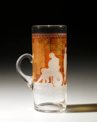
W. L. Libbey & Son, New England Glass Works
Lemonade Glass (Etched With "The Departure Of Briseis From The Tent Of Achilles" After John Flaxman)
1884